 | ÐлекÑÑоннÑй компоненÑ: A701 | СкаÑаÑÑ:  PDF PDF  ZIP ZIP |
Äîêóìåíòàöèÿ è îïèñàíèÿ www.docs.chipfind.ru
Specification :
·Specimen : Paraformaldehyde-fixed, paraffin-embedded 1.0mm diameter 12
different types of cancer tissue and matching normal tissue cores. Single spot
for each tissue type.
·Packing status :
Each array slide is individually packed in i) a hard plastic
case and ii) an opaque aluminum bag sealed under a nitrogen atmosphere to
prevent oxidation and drying.
·Enclosed documents:
product specification (specification, layout,
coordinates of tissue spots), H&E stained images, general protocols (3
pages).
·3.5 inch diskette :
This contains a data set related to the tissues on the
slide, in MS Excel format.
Storage and handling
Shipped at room temperature. Recommended storage conditions upon arrival:
2-8
. Each individually packed aluminum foil envelope has been filled with
nitrogen gas. For maximum antigenicity, use the slide as soon as possible
after opening.
For research use only
(Paraformaldehyde fixed)
A701: Test slide, various cancers plus
corresponding normal
PETA
TM
Array
TEST SLIDE
Section No.
1 2
3
4
5
6
A
B
C
D
A
B
C
D
Label your own
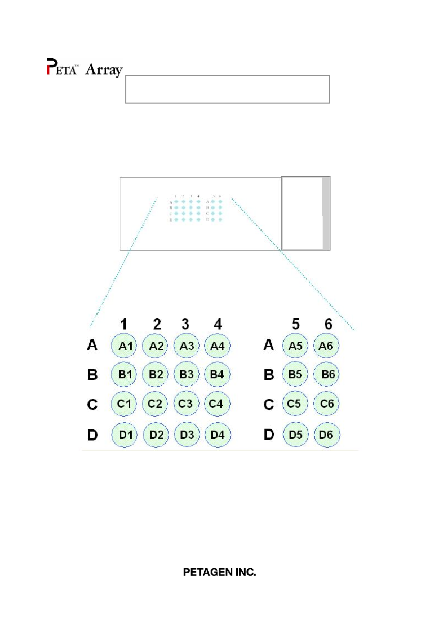
Layout
For research use only
A701: Test slide, various cancers plus
corresponding normal
(Paraformaldehyde fixed)
P
E
T
A
T
M
A
r
r
a
y
T
E
S
T
S
L
I
D
E
S
e
c
t
i
o
n
N
o
.

For research use only
(Paraformaldehyde fixed)
Coordinates of tissue spot
A701: Test slide, various cancers plus
corresponding normal
No.
Age Sex
Tissue Type
Pathology diagnosis
1 A
1
29
m
Skin
Squamous cell carcinoma
2 B
1
29
m
Skin
Normal
3 A
2
48
f
Breast
Ductal carcinoma
4 B
2
48
f
Breast
Normal
5 A
3
32
f
Thyroid gland
Follicular carcinoma
6 B
3
32
f
Thyroid gland
Normal
7 A
4
69
m
Lung
Squamous cell carcinoma
8 B
4
69
m
Lung
Normal
9 A
5
29
f
Liver
Hepatoma
10 B
5
29
f
Liver
Normal
11 A
6
58
f
Kidney
Renal cell carcinoma
12 B
6
58
f
Kidney
Normal
13 C
1
45
m
Bladder
Transitional cell carcinoma
14 D
1
45
m
Bladder
Normal
15 C
2
34
f
Ovary
Serous carcinoma
16 D
2
34
f
Ovary
Fallopian tube
17 C
3
33
f
Uterus
Cervix carcinoma
18 D
3
33
f
Uterus
Normal
19 C
4
64
m
Esophagus
Squamous cell carcinorma
20 D
4
64
m
Esophagus
Normal
21 C
5
67
m
Stomach
Signet ring cell carcinoma
22 D
5
67
m
Stomach
Normal
23 C
6
70
m
Colon
Adenocarcinoma
24 D
6
70
m
Colon
Normal
Coordinate

For research use only
A701: Test slide, various cancers plus
corresponding normal
(Paraformaldehyde fixed)
A1 A2 A3 A4
A5 A6 B1 B2
B3 B4 B5 B6
C1 C2 C3 C4
C5 C6 D1 D2
D3 D4 D5 D6
QC sheet_LOT#13101200304191
Haematoxylin and Eosin staining

No. Age
Sex
Specimen
Key word
Pathological information
A1
B1*
m
29
skin
invasive squamous
cell carcinoma
Skin, scalp, excision:
1. Invasive squamous cell carcinoma, well-differentiated with extension to the
subcutaneous fat tissue (invasion depth: about 3cm).
2. Resection margins, all circumference and base: Free of tumor.
3. Lymph nodes, level 2a(0/36), level 2b(0/11), level III(0/25), level IV(0/13) and level
V(0/11):(0/96): Free of tumor.
A2
B2*
f
48
breast
infiltrating ductal
carcinoma
Breast, right, modified radical mastectomy:
1. Infiltrating ductal carcinoma
1) Black's nuclear grade 1(poorly differentiated).
2) Modified Bloom and Richardson's histological grade III (tubule formation:3, nuclear
pleomorphism:3, mitosis:2).
3) No vascular invasion.
2. Regional lymph node, axillary(0/18), level III(0/5):(0/23): Free of tumor.
Note: ER(+), PR(+), C erb B2(-).
A3
B3*
f
32
thyroid
follicular
carcinoma
Thyroid gland, right, thyroidectomy:
Follicular carcinoma, minimally invasive, showing capsular invasion.
A4
B4*
m
69
lung
1.squamous cell
carcinoma
2.obstructive
pneumonitis
3.pneumonia
Lung, right, pneumonectomy:
1. Upper lobe: Squamous cell carcinoma, moderately differentiated,
with - extension to the visceral pleura.
- marked central necrosis.
- tumor size: 6.3x5.5cm.
- obstructive pneumonitis in the remaining lung.
2. Regional lymph nodes: Free of tumor in all nodes(0/13), in detail, subcarinal(0/6), lower
paratracheal(0/2), regional(0/5).
A701 : test slide, various cancers + corresponding normal tissues

No. Age
Sex
Specimen
Key word
Pathological information
A701 : test slide, various cancers + corresponding normal tissues
A5
B5*
f
29
liver
hepatocellular
carcinoma
Liver, right, lobectomy:
Hepatocellular carcinoma,
with 1) size: 8x7.6x7.5cm
2) Edmondson grade II
3) macrotrabecular and pseudoglandular types
4) infiltrative type
5) capsular invasion
6) necrosis: less than 5% of total volume
7) portal vein invasion
8) intact hepatic resection margin
9) Non-neoplastic liver showing congestion
A6
B6*
f
58
kidney
renal cell
carcinoma
Kidney, left, radical nephrectomy:
1. Renal cell carcinoma, conventional(clear cell) type, with
1) Fuhrman's nuclear grade: 4
2) Foci of sarcomatoid differentiation
3) Invasion to the perinephric fat tissue but not to Gerota's fascia
4) Renal vein involvement (pT3b)
2. Lymph nodes, perihilar(0/11), paraaortic(0/2) and left common iliac(0/3): Free of tumor
metastasis in all 16 nodes.
C1
D1*
m
45
bladder
urothelial
carcinoma
Urinary bladder including prostate, seminal vesicle and ureter, radical cystectomy:
Urinary bladder: Papillary urothelial carcinoma, high grade, with squamous differentiation,
with various pathologic state including low grade papillary urothelial carcinoma, urothelial
tumor of low malignant potential, carcinoma in situ and papilloma, with extension to the
perivesical soft tissue and prostate, and with extensive lymphatic, venous and perineural
invasion, incompletely excised.
C2
D2*
f
34
ovary
serous tumor
borderline
malignancy
Ovary and fallopian tube, side unstated, salpingooophorectomy:
Ovary: Borderline serous and mucinous tumor with serous micropapillary pattern (so called
micropapillary serous adenocarcinoma by Kurman), confined within ovarian capsule.
The matched normal tissue of this case is fallopian tube, not ovary.

No. Age
Sex
Specimen
Key word
Pathological information
A701 : test slide, various cancers + corresponding normal tissues
C3
D3*
f
33
uterus(cervix)
invasive squamous
cell carcinoma
Cervix: Invasive squamous cell carcinoma, large cell, keratinizing (invasion depth: 1.1cm)
with focal lymphovascular permeation
C4
D4*
m
64
esophagus
basaloid
carcinoma
Esophagus, esophagectomy:
Basaloid squamous cell carcinoma
with 1) size: 2.7x2.0x2.0cm.
2) expanding growth.
3) involvement at submucosal space and extension to upper border of proper muscle
layer.
4) intact proximal and distal resection margin.
5) no tumor metastasis to upper paraesophageal lymph node (separately
submitted:0/2)
C5
D5*
m
67
stomach
signet ring cell
carcinoma
Stomach, subtotal gastrectomy:
1..Signet ring cell carcinoma
1. Diffuse infiltrative type
2. With extension to serosa and perigastric fat tissue(SE)
3. Frequent lymphatic permeation and perineural invasion
4. Focally mixed with tubular adenocarcinoma, moderately differentiated
5. Mixed type by Lauren's classification and infiltrative type by Ming's classification
2.. Regional lymph nodes, No.3(6/5), No.4(1/1), No.5(0/0), No.6(9/14), No.7(1/9),
No.8(0/3), No.12(0/1), No.13(0/3), No.17(0/2):(17/38): Tumor metastasis in 17 out of 38
nodes.
C6
D6*
m
70
colon
adenocarcinoma
1.Sigmoid colon, radial sigmoid colectomy:
A. Adenocarcinoma, poorly differentiated, ulceroinfiltrative type with extension to
pericolic fat tissue and is very close to lateral margin (about 0.5mm).
B. Tubular adenoma with high grade dysplasia.
Resection margins, proximal and distal: Free of tumor.
2. Regional lymph nodes, principal(0/7), pericolic(2/22):(2/29): Tumor metastasis in 2 out
of 29 lymph nodes.
*: corresponding normal tissues

Appendix: application protocol 1
Deparaffinizing and H&E stain
For research use only
Deparaffinization and hydration
Dry the slide at 58 for 1hr or overnight
, before deparaffinization (put slides in horizontal position)
Xylene (removal of paraffin) 4 X 10 min
100% Ethanol (de xylene)
95% Ethanol 1min
95% Ethanol 1min
80% Ethanol 1min
70% Ethanol 1min
Wash (tap water) until washing is completed (5 min)
Routine H&E stain
Wash (tap water) until washing is completed (5 min)
Hematoxylin (Harris,nucleus staining,over staining) 3 min
Wash (tap water)
Decolor in 0.1% HCl, 70% Ethanol : repetitive dipping
Neutralization 10 min (top water 5min/ Ammonia water repetitive dipping)
Eosin (cytoplasm staining) 1 min
Washing 30 sec
70% Ethanol 1 min
80% Ethanol 1 min
95% Ethanol 1 min
95% Ethanol 1 min
100% Ethanol 1 min
Xylene (clear to increase refractive index to 1.5 fold) 4 X 10 min
Mount with mounting solution ( eg. balsam)

Appendix: application protocol 2
Immunohistochemistry
For research use only
IHC(immunohistochemistry)
Immunohistochemistry is an exquisitely sensitive method for locating an antigen within a cell or tissue through a
high-resolution image (a single cell among thousands or millions). The method is based on the use of a primary
antibody binding specifically to its cognate antigen. The bound antibody is then visualized by colorimetric or
fluorescent detection methods.
IHC procedure
The protocol needs to be optimized for antibody you
The protocol needs to be optimized for antibody you
may want to test and/or you might need to follow
may want to test and/or you might need to follow
instructions from suppliers.
instructions from suppliers.
Antigen retrieval method
During the preparation of tissues for staining,
During the preparation of tissues for staining,
antigens are heavily modified by the fixatives
antigens are heavily modified by the fixatives
frequently on free amino acid groups. Because they
frequently on free amino acid groups. Because they
can be hidden by other molecules, antigen retrieval
can be hidden by other molecules, antigen retrieval
procedure is required to counter these changes.
procedure is required to counter these changes.
There are several methods for antigen retrieval. The
There are several methods for antigen retrieval. The
selection is made according to the experimental
selection is made according to the experimental
purposes. If the experiment is the conditioning
purposes. If the experiment is the conditioning
process with first trial of that antibody, various
process with first trial of that antibody, various
methods need to be tried.
methods need to be tried.
Dry a slide at 58 overnight
Deparaffinize in xylene
Hydrate the slide in gradient ethanol
Retrieve antigen (see left section)
Dip in 3% H
2
O
2
10-15 min and washing buffer
3 X 5 min
Block with normal serum
1. Direct method
Biotin-tagged Primary Ab for 1~2 hr at RT or
37 incubator or for overnight at 4(don't
wash, just change the blocking solution for
primary antibody) and washing buffer 2 X 5 min
2. Indirect method
-1. Biotin-tagged primary Ab for 1~2 hr at RT or
37 incubator or for overnight at 4(don't
wash, just change the blocking solution for
primary antibody) and washing buffer 2 X 5 min
-2. Biotin-tagged secondary Ab for 10-15min at
RT and washing buffer 2 X 5 min
ABC reagent (streptavidin-HRP) for 10~15min
and washing buffer for 2~3 X 5 min
Fresh chromogen (DAB or AEC) for 1~3 min and
Wash with tap water
Counter stain (the nucleus) with Hematoxylin
or methyl green
-1. When Hematoxylin is used :
dehydrate in gradient ethanol (70% to 100%)
and clear with xylene,
mount with insoluble mounting medium
(eg. balsam)
-2. When methyl green is used : just dry and mount
with soluble mounting medium (eg. glycerin or
gelatin)
-3. When AEC is used for chromogen :
just dry and mount with soluble mounting medium
1. Proteolytic enzyme pre-treatment method
Cleave the bonds formed from the fixation process.
Cleave the bonds formed from the fixation process.
Enzymes routinely used include
Enzymes routinely used include
trypsin
trypsin
,
,
pronase
pronase
or
or
pepsin.The concentration and reaction time must be
pepsin.The concentration and reaction time must be
controlled since the excess enzyme treatment can
controlled since the excess enzyme treatment can
damage the target antigens.
damage the target antigens.
- pronase: 0.05%(W/V) in PBS or
- trypsin : 0.05%(V/V) in PBS
- pepsin : 0.05%(V/V) in 2N HCl
Incubate in one of the above solutions
at RT or 37 for 18 min
Dip in cold DW
2. Heat-induced antigen retrieval method
Antigens fixed in formalin are hidden by fixative and
Antigens fixed in formalin are hidden by fixative and
calcium ions. Chelating or precipitating these
calcium ions. Chelating or precipitating these
calcium ions by specific solutions like citrate buffer,
calcium ions by specific solutions like citrate buffer,
EDTA and EGTA with heating can cleave these
EDTA and EGTA with heating can cleave these
bonds.
bonds.
Place the slides into a rack
Immerse the slides in citrate buffer*
Move the entire container into microwave oven
Microwave the slides at maximum watt for 4 X 5 min
( after each cycle, replenish any lost liquid from
the slide container by addition of DW)
Remove the container and allow it to cool to RT
Wash with appropriate washing buffer
*citrate buffer : 0.01M citric acid, pH 6.0

IHC Conditioning
Negative controls
without a primary antibody, without a secondary antibody, or without detecting reagents.
Why does my negative control show strong
signal?
The signal is due to non-specific cross-reactivity of
detection reagents.
Dilute the secondary Ab.
Change species of secondary Ab.
Detection reagent only is omitted. Intrinsic tissue
enzyme activity is interfering with the reaction. Treat
tissue with reaction solution